Posts

Showing posts with the label Biology

What is Homeostasis, Osmoregulation and Excretion?

  Homeostasis - homeo means same and stasis means standing. The definition of homeostasis is maintenance of the constant internal environment of the body. Internal environment is very necessary for normal life processes. The osmoregulation, excretion and homeostasis are inter linked with each other. Osmoregulation and excretion help in maintaining chemical and fluid homeostasis.  Osmoregulation- It is the process in which they regulate the body salt and water content. This process maintain the composition of the body fluids at steady state for efficient metabolism in the cells. The body fluids are of two types- i) Intracellular fluid- Found within the cells. ii) Extracellular Fluid - Found outside the cells.  Excretion - It regulates the volume, composition, ionic contents, pH and osmotic pressure of the body fluid by removing substance in harmful quantity and conserving material necessary for normal functioning of the body. Excretion maintain the homeostasis. All body fluids exchang
Body Fluids and Circulation?

  Blood and Lymph are the two body fluids which constant supply the useful materials and simultaneously remove the waste substances. The main materials that need to transport in the body is gases, nutrients, hormones, waste materials and molecules. Blood and Lymph fluids are used by most of the higher organism. Circulation of blood fluids is also known as circulatory system.  Fluid is circulate inside the body by Intracellular circulation and Extracellular circulation.  Intracellular circulation- It occur in unicellular organisms. All the substances are distributed by regular streaming movement. It also brings carbon dioxide and nitrogenous waste near the cell membrane. Paramecium, Amoeba are the example of intracellular circulation.  Extracellular Circulation - some animals have additional techniques of fluid circulation outside the cell.  1) Environmental fluid circulation - Some invertebrates have no circulatory fluid. These animal circulate the surrounded water through the caviti

The Human Heart? Structure, Working, Heart beat, Heart rate, Heart Sound and Cardiovascular diseases.

Image
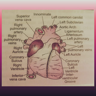
Human heart is the hollow muscular organ of the body which pump and supply the blood throughout the circulatory system. It is located a little to the left of the middle of chest. Heart is very sensitive organ and more responsible for life, so it covered with the rib cage to protect any injury. The weight of heart is about 240 grams and the size of heart is about a fist.  Structure -   The structure of heart is divided into two parts- Exterior structure and Interior structure.  1) Exterior structure -  Heart is made up of three layers of tissue- Pericardium, Myocardium and Endocardium.  i) Pericardium - is the outer layer of heart. It is made up of two layers Outer parietal pericardium and inner visceral pericardium. Between these layer their are potential space called pericardial cavity. This Pericardial cavity contain up to 50ml of pericardial fluid. This fluid keeps the heart moist and reduce the friction between heart wall and surrounding tissue, when the heart beats.  ii) Myocardi

Difference between Arteries and Veins?

Image
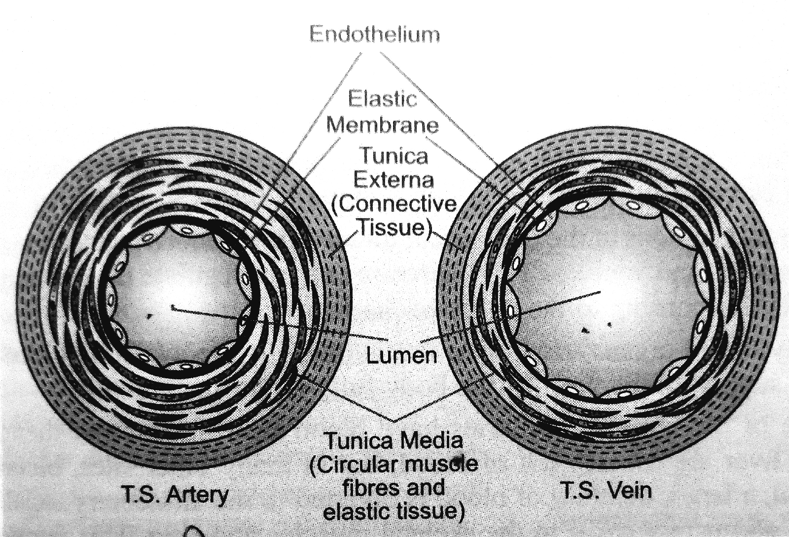
Arteries 1) Arteries distributes blood from Heart to Body.  2) The flow of blood is fast due to heart beats. 3)  Arteries contain aerated blood. 4) In arteries their are no valves present in it. 5) Arteries become empty after the death of organism.  6) The tunica media is thin, because it contains fewer muscle fibers and elastic fibers.  7) Lumen in arteries are narrow. Vein 1 ) Veins brings the blood from Body to Heart. 2)  The flow of blood is slow.  3) Veins contain non-aerated blood.  4)  Veins contain valves, to prevent back flow of blood.  5) Blood is still in vein after death of organism.  6)  The tunica media is thicker than arteries, because it contain more muscle fibres and elastic fibres.  7) Lumen in veins are wider than arteries. 

Difference between Open Circulatory system and Closed Circulatory System.

Image
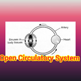
                                                 Open Circulatory System 1) In this the blood is direct contact with the tissue cells. 2) Exchange of material occur directly between the blood and tissue cells.  3) Blood flow by sinuses without proper walls. 4) In open circulatory system blood has very low pressure. 5) The flow of blood is very slow. 6) Blood flow cannot be regulated, so that this system is less efficient. 7) Because of no red cells, the respiratory pigment dissolved in the plasma of blood.  8) Open circulatory system found in Prawns, crabs, spiders, insects and most mollusks like snails.                              Closed Circulatory System 1) In this the blood is not direct contact with the tissue cells.  2) Exchange of material between tissue and blood occur via tissue fluid. 3) Blood flow by blood vessels with proper walls. 4) In this system the blood has high pressure. 5) The flow of blood is high.  6) Blood flow can be regulate, so that this system is more effici

Human Respiratory System? Explained.

Image
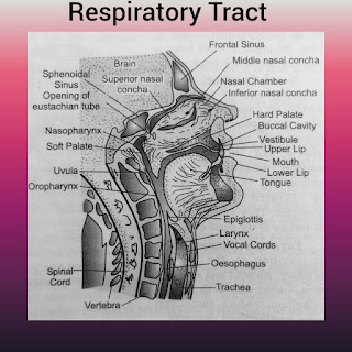
  Human Respiratory system is a part of body which provide a way for breathing and surface for gases exchanges. Body get oxygen through respiratory system. It is divide into two parts-  1) Respiratory Tract 2) Respiratory Organ 1) Respiratory Tract - Tract means way. Respiratory tract is a passage way for the fresh air to come inside lungs and foul air to  pass away from lungs to environment. It consist nostrils, nasal chamber, internal nares, pharynx, larynx, trachea, bronchi and bronchioles. i) Nostrils - known as external nares. A pair of slits at the lower end of the nose. They open into the next way that is nasal chamber.  ii) Nasal chamber - It is a pair of passages in the head located above the palate. By nasal septum, both chamber are separate from each other. These chambers shows three regions-              Vestibular Region - It lies just within the external naris. This part is very short and lined with skin. It bear hairs, some sweet and oil glands. The function of hairs i

WHAT IS RESPIRATION? Its TYPES and FUNCTIONS?

Image
          Respiration is the intracellular biological process occur in all organisms. It involving the exchange of environmental oxygen with carbon dioxide that produced inside the cell during oxidation of food .  Organism take the oxygen from environment. Carbon dioxide release during oxidation and eliminate to the environment. While oxidation of food, small amount of energy release and this energy stored in the form of ATP.  This ATP hydrolyze into ADP when body required energy. The surface at which gases exchange known as respiratory surface. Types of respiration -  Respiration are two types Aerobic respiration and Anaerobic respiration 1) Aerobic Respiration - means with oxygen. It is the process in which the molecular oxygen utilize and carbon dioxide release simultaneously. It yields inorganic end product but  provide much more energy. It is found in animal and plants.   Aerobic respiration is divide into two types -  Direct Aerobic Respiration and Indirect Aerobic respiration. 

What is Cell division and its types?

Image
Cell Division - Cell division is a process in which the single cell divide or multiply into two or more identical cells. It play important role in growth, reproduction and repair. There are three types of cell division-  1) Amitosis 2) Mitosis 3) Meiosis 1) Amitosis - is the process in which the cell is directly divide into two daughter cell without any formation of spindle and appearance of chromosomes. The nucleus present inside the cell elongates and develops a constriction round its middle. This constriction gradually deepens and finally cuts the nucleus into two daughter cells. Example - Paramecium divide by amitosis.   2) Mitosis- is the process in which two identical daughter cell produced by a single parent cell by karyo-kinesis, cytokinesis and cell separation. The daughter cells carry same amount of DNA, same amount and kind of Chromosomes and same hereditary instruction from parent cell so that this division also known as equational division. It occur in all kind of eukary

What is MITOSIS?

Image
MITOSIS is the one of cell division method. It is the process in which the parent cell divide into two identical daughter cells. Both daughter cells having nucleus with same amount of DNA, chromosomes and same hereditary instructions from parent cell. This division called equational division. It takes place in somatic cells of animals. So that it also known as somatic division.  In 1875 Eduard Strasburger   firstly described the mitosis in plant cells. After that, In 1879 Walther Flemming described the mitosis in animal cells.  Mechanism of Mitosis- The parent cell divide into two equal daughter cells by Karyo-kinesis, Cytokinesis and Cell separation. Karyo-kinesis is the division of  nucleus. Cytokinesis is the division of  cytoplasm. The duration of Mitosis is from 30 minutes to 3 hours.  A. Karyo-kinesis  is the division of cell nucleus. This process divide into four stages-  1) Prophase 2) Metaphase 3) Anaphase  4) Telophase 1) Prophase - It is the starting phase of mitosis wher

Difference between Osmosis and Diffusion?

Image
                                                                                                                                       Osmosis - 1) It is the movement of solvent from higher water concentration to lower water concentration . 2) It take place through semi permeable membrane. 3) Osmosis occurs in liquid medium only.    4) Only the solvent molecules move from one place to another. 5) It does not equalize the concentration of solvent molecules. 6) Osmosis can be stopped and reversed by applying external pressure. 7) Example - Sugar molecules are solute and the water molecules are solvent. This example shows the movement of water molecules (solvent) from higher concentration to lower concentration. Diffusion - 1) It is the movement of solute from higher concentration to the lower concentration. 2) This process does not require semi- permeable membrane. 3) Diffusion occur in any medium solid, liquid

BLOOD GROUP?

Image
Blood is a circulating life sustaining fluid. It provides oxygen and nutrients for cells, tissue and organs. It keeps body warm 37degreeC. In some clinical cases we need to do blood transfusion. The blood transfusion is done on the basis of BLOOD GROUP.  Blood are four types A, B, AB, O these are known as ABO blood group . The types of blood group is depend upon the presence and absence of antigen and antibody on Red blood cells. In 1900 Karl Landsteiner recognized these types of blood group in human being. He observed that ABO blood group are determined by gene I. This gene I have three alleles IA, IB and IO.  Each blood group has antigen on the surface of red blood cell and opposing antibody in plasma.  The presence of antigen and anti-antibody accordingly-  1) Blood group A - In this the A antigen is present on the surface of red blood cell and Anti-B antibody present in plasma 2) Blood Group B - In this the B antigen present on the surface of red blood cell and anti-A antibody pre